Amyloidogenecity of Epidermal proteins: Unfolding the mysteries.
| Image via Wikipedia |
[This is a paper on cutaneous amyloidosis that I did not publish. I presented it in a conference though. The discussion is posted in my dermatology blog. Poster can be downloaded here: http://www.box.com/s/uae8yijuohefphzoh4cx under 
Amyloidogenecity of Epidermal proteins by Bell Raj Eapen is licensed under a Creative Commons Attribution-ShareAlike 3.0 Unported License.
Introduction
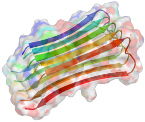
Amyloidosis is the process by which biologically unrelated proteins fold in a non-native way thereby acquiring certain novel but generic properties. Amyloid cannot be always classified as a misfolded protein as amyloid deposits can have physiological properties. Ultrastructurally amyloid has antiparallel, beta-pleated sheet configuration. Amyloid deposits occur in several organs including skin. Cutaneous amyloidosis occurs in two main patterns called macular and lichen amyloidosis. The discernible properties of amyloid important for dermatologists include apple-green birefringence under polarizing light and congo-red staining. Since several unrelated proteins can form amyloid deposits, it is not easy to identify the source of amyloid deposits. Since cutaneous amyloid deposits bind to antikeratin antibodies it is presumed to be a keratin derivative. However cutaneous amyloidosis being innocuous has not received as much attention as neuro-degenerative disorders.
The propensity for a protein to form amyloid is determined by short segments of amyloidogenic fragments composed mostly of either hydrophobic residues or a combination of Gln and Asn residues. Extrinsic factors like pH and the concentration of polypeptide may also influence rate of aggregation. Several computational methods for amyloidogenic fragments are described. Though keratin is widely accepted as the source of amyloid in cutaneous amyloidosis, the probable involvement of other proteins like collagen and elastin is suspected but not proved. We have used a web-based ‘FoldAmyloid server’ to predict amyloidogenic regions on these proteins to have a better understanding of their behaviour. The FoldAmyloid algorithm utilizes the expected probability of hydrogen bonds formation and expected packing density of residues to predict amyloidogenic regions on a protein and has shown good success rates in predicting amyloidogenic regions on polypeptides. We have also analysed V324A mutation on KRT5 in a Weber Cockayne variant of Epidermolysis Bullosa reported to be associated with amyloid deposits. We also discuss the probable molecular mechanisms of common but ill-explained symptoms of cutaneous amyloidosis like pruritus and hyperpigmentation. Finally we analyse the mechanism of action of few existing treatment strategies and the future perspectives.
Materials and Methods
The web based FoldAmyloid server was used to predict the amyloidogenic regions on keratin, collagen and elastin. FoldAmyloid server does not support batch submission of multiple sequences. Hence a perl script was written to retrieve specified sequences from GenBank and submit the sequences one by one to FoldAmyloid server. All available isoforms of Keratins 1 to 20, collagen 1 to 25 and elastin were submitted for prediction of amyloidogenic areas using the script. The rare mutation, V324A on KRT 5 reported to be associated with amyloid deposits was submitted separately. The default settings of foldAmyloid were used for prediction.
Related articles
- Formation of immunoglobulin light chain amyloid oligomers in primary cutaneous nodular amyloidosis (dx.doi.org)
- Noncore Residues Influence the Kinetics of Functional TTR(105-)(115)-Based Amyloid Fibril Assembly. (ncbi.nlm.nih.gov)
- Slow Amyloid Nucleation via alpha-Helix-Rich Oligomeric Intermediates in Short Polyglutamine-Containing Huntingtin Fragments. (ncbi.nlm.nih.gov)
- Inhibiting the Nucleation of Amyloid Structure in a Huntingtin Fragment by Targeting alpha-Helix-Rich Oligomeric Intermediates. (ncbi.nlm.nih.gov)

